Cranial Cruciate Ligament Disease in Dogs
Cranial cruciate ligament (CCL) disease is one of the most common causes of hindlimb lameness and pain in dogs, and is similar to an ACL injury in humans. In dogs, there are two main ligaments that stabilize the knee joint (also called the stifle joint). There is the cranial cruciate ligament (CCL) that extends from the front of the femur bone and spans to the back top of the tibia, and the caudal cruciate ligament, which starts on the back of the femur and extends to the front of the tibia. The purpose of the CCL is to stabilize rotation of the joint and to prevent forward sliding of the tibia. Dogs can have either a partial or complete tear of the ligament, often resulting in extreme pain and traumatic injury. Commonly, the ligament degenerates over time following injury, ultimately resulting in complete failure of the ligament.
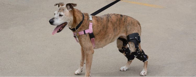
Image provided by the Animal Medical Center.
Predisposed Breeds
Any breed of dog can suffer from a cruciate injury, but it is typically seen in large-breed dogs.Genetic inheritance of CCL disease has been specifically noted in Newfoundland and Labrador Retrievers, but also affects Boxers, Rottweilers, Staffordshire Terriers, Akitas, and Mastiffs.
Risk Factors
The top risk factors that predispose a dog to developing a CCL injury are obesity and early neutering. Obesity causes strain and is thought to increase the load on the joints, leading to injury. Early neutering has a direct pathway to CCL injury.
Two theories have been postulated for early neutering and its influence on CCL injury. The first states that sex hormones influence the ligament and early disruption of the hormones affects the strength of the ligament. The second theory is that early neutering could alter tibial and femoral growth plate closure. Other risk factors that may affect the CCL and have been associated with the likelihood of degeneration are advanced age, excessively straight hindlimb conformation, and immune-mediated joint diseases.
Clinical Signs
Clinical signs of CCL injuries are dependent on a few variables, including the chronicity of injury and whether the tear is partial or complete.
The most common signs of a CCL injury are:
- Acute, non-weight bearing lameness
- Pain when placing weight on the leg
- Thickening of the medial (inside of the joint), also known as medial buttress
- Reluctance to rise/stand
- Reduced activity
Lameness is a sign of pain and could be secondary to a cruciate injury, as well as other injuries like arthritis, patella luxation (medial or lateral), hip dysplasia, hip luxation, fracture or broken bones, cancer, joint diseases (arthropathy), and meniscal injury. If your dog is limping, they should be evaluated by your veterinarian. During the veterinarian’s physical exam, they will be able to rule out other causes of lameness and make recommendations on the next diagnostic steps.
Diagnosis
Diagnostics testing for cruciate injury includes:
- A complete medical history. This helps a veterinarian determine the chronicity of the problem and isolate patterns if present.
- A physical exam. A physical exam allows a veterinarian to isolate which leg and joint is affected. In dogs with chronic cruciate injuries, scar tissue develops on the medial (inside) of the affected knee, which is easily palpated during an exam and is called a medial buttress. There are two tests that a veterinarian can perform when palpating a dog’s knee to diagnose cruciate injury.
- Cranial drawer sign. This test involves the veterinarian trying to elicit forward movement of the tibia (shin bone). The cranial ligament is designed to prevent this forward movement.
- Tibial thrust. This test involves extending the hind leg fully and flexing the toes. If pain is noted, cruciate injury is probable.
- Radiographs. Radiographs of the limb can help confirm a cruciate injury and rule out other diseases like fracture, luxation, and cancer. Findings on orthopedic radiographs that are supportive of a cruciate injury are:
- Joint effusion. Fluid in the knee joint.
- Osteophyte formation. New bone formation within the joint. This is commonly seen with underlying arthritis.
- CT scan or MRI. In complicated cases, further imaging by CT scan or MRI is recommended.
Treatment Options
Once a diagnosis of a cruciate injury has been made, treatment options can be discussed. Surgical options are generally recommended, and are the only way to stabilize the joint and minimize arthritis formation. Medical therapy is also an option for dogs that are not candidates for surgery, due to other underlying diseases, age, or financial constraints.
Both surgical and medical treatment options rely on exercise restriction, pain control, joint supplements, and weight loss (if applicable).
- Exercise restriction is important to minimize pain and inflammation. In both medically and surgically managed cruciate injuries, exercise is restricted for 4-6 weeks. This means no running, jumping, playing, leashed walks, or outdoor bathroom use.
- Pain control is usually achieved through veterinary, non-steroidal, anti-inflammatory medications, as well as other medications based on severity. Your veterinarian will discuss which medication is best for your dog and perform blood tests before prescribing medications. DO NOT administer human pain medications to your dog without a veterinarian’s advice.
- Glucosamine, chondroitin, and omega 3 fatty acids are the most commonly recommended joint supplements. Your veterinarian can suggest veterinary-formulated products or give you the appropriate dose to give in a human joint supplement.
- Weight loss is extremely important in medically and surgically managed patients. Weight loss can be difficult in exercise-restricted dogs, therefore, counting calories and conservatively reducing caloric intake is the most successful weight loss strategy. Your veterinarian can help you formulate a healthy weight loss plan for your dog.
Medical Therapy
Medical therapy for cruciate disease focuses on the above treatment options in addition to physical rehabilitation, often involving underwater treadmill or swimming exercises. There are also braces and orthopedic support devices marketed to help stabilize the joint during the healing process. Your veterinarian can work with you to create a treatment plan and determine if medical therapy is right for your dog.
Surgical Therapy
Surgical therapy is currently the recommended therapy for dogs with cruciate disease. There are multiple surgical techniques based on veterinarian preference, referral to boarded veterinary surgeon, budget, and size of dog. No particular technique is best for every dog, therefore, it is important to discuss available options with the veterinarian performing the surgery.
Available surgical options are divided into two groups: suture-based techniques and osteotomy-based techniques:
- Suture-based techniques. These techniques use a heavy grade suture, either outside the joint or inside the joint, to mimic the job of the cranial cruciate ligament. These techniques are usually less expensive than osteotomy-based surgeries. They are more successful in small dogs due to risk of complication in large-breed and highly active dogs.
- Extracapsular suture stabilization is the most common suture-based surgery performed.
- Intracapsular suture stabilization also known as the “tightrope” technique is less commonly used.
- Osteotomy-based techniques. These surgeries involve making an incision into a bone (osteotomy) and using implants (metal plates) to change the biomechanics of the joint instead of recreating the ligament. These techniques are usually recommended for active medium to large-breed dogs. Two techniques are currently being used and are based on surgeon’s preference, experience, and training. Osteotomy-based techniques require advanced training and are usually done by boarded veterinary surgeons. The two techniques are:
- Tibial Plateau Leveling Osteotomy (TPLO)
- Tibial Tuberosity Advancement (TTA)
Long-term recovery is unique for each dog. 85-90% of dogs that undergo surgical stabilization of the joint have significant improvement in mobility after the initial recovery phase. The veterinarian involved in your dog’s surgery will give detailed recovery recommendations which should be strictly followed. After osteotomy-based surgery, strict rest is required to let the bone heal, and radiographs should be taken before exercise is resumed. Unfortunately, 40-60% of dogs that have a cruciate injury to one knee will have a similar injury to the opposite knee within the subsequent year. Weight loss and physical rehabilitation can help during the postoperative phase and when exercise is reintroduced.